
KPV modulates inflammatory signaling in studied cellular systems through intracellular effects associated with reduced NF-κB activation; PepT1-mediated uptake is an important part of the published intestinal-cell evidence. KPV is not interchangeable with its parent peptide, α-MSH, and those findings do not establish a universal receptor mechanism. This guide examines the molecular distinction, experimental evidence, and analytical requirements strictly for research use.
- The KPV peptide mechanism connects PepT1-mediated cellular uptake with reduced NF-κB signaling in published intestinal-cell experiments.
- KPV contains three amino acid residues; shared sequence with α-MSH does not establish shared receptor pharmacology.
- 4-Amino-Labs research-chemical sourcing belongs alongside batch identity checks, not assumptions about biological activity.
- Separate peptide uptake, pathway readouts, and inflammatory markers when interpreting mechanism evidence.
Contents
- How does KPV modulate inflammatory signaling in research?
- Why the molecular distinction matters
- What PepT1 uptake establishes
- What NF-κB measurements establish
- Why KPV findings vary
- How analytical documentation supports interpretation
- Related mechanism questions
- Limitations of the evidence
- FAQ
- One last thing
- References
- Related guides
How does KPV modulate inflammatory signaling in research?
The clearest mechanistic distinction is between cellular entry and downstream signaling: PepT1 transports KPV, whereas NF-κB measurements describe a subsequent biological response. Dalmasso and colleagues reported PepT1-mediated KPV uptake and inhibition of inflammatory signaling in intestinal epithelial cell experiments. Their findings connect transport with activity in the systems examined; they do not identify PepT1 as an inflammatory signaling receptor. 1
For your 2026 literature review, separate the evidence into these levels:
| Evidence level | Finding or measurement | Supported interpretation | Interpretation to avoid |
|---|---|---|---|
| Molecular identity | Lysine–proline–valine sequence | Defines the tripeptide under discussion | Every related fragment has equivalent activity |
| Cellular entry | PepT1-mediated transport | Transport contributes to intracellular access in studied cells | PepT1 is the peptide's signaling receptor |
| Pathway activity | Reduced NF-κB activation | An inflammatory transcriptional pathway changes | A direct molecular binding target is established |
| Downstream response | Inflammatory mediator measurements | Supports a functional cellular response | Every inflammatory pathway is inhibited |
Material characterization belongs alongside those distinctions. A useful starting point is the guide to verifying peptide purity with third-party COA testing, because an interpretable mechanism claim starts with an identified research material.
Why the molecular distinction matters
KPV is a fragment, not a shorter equivalent
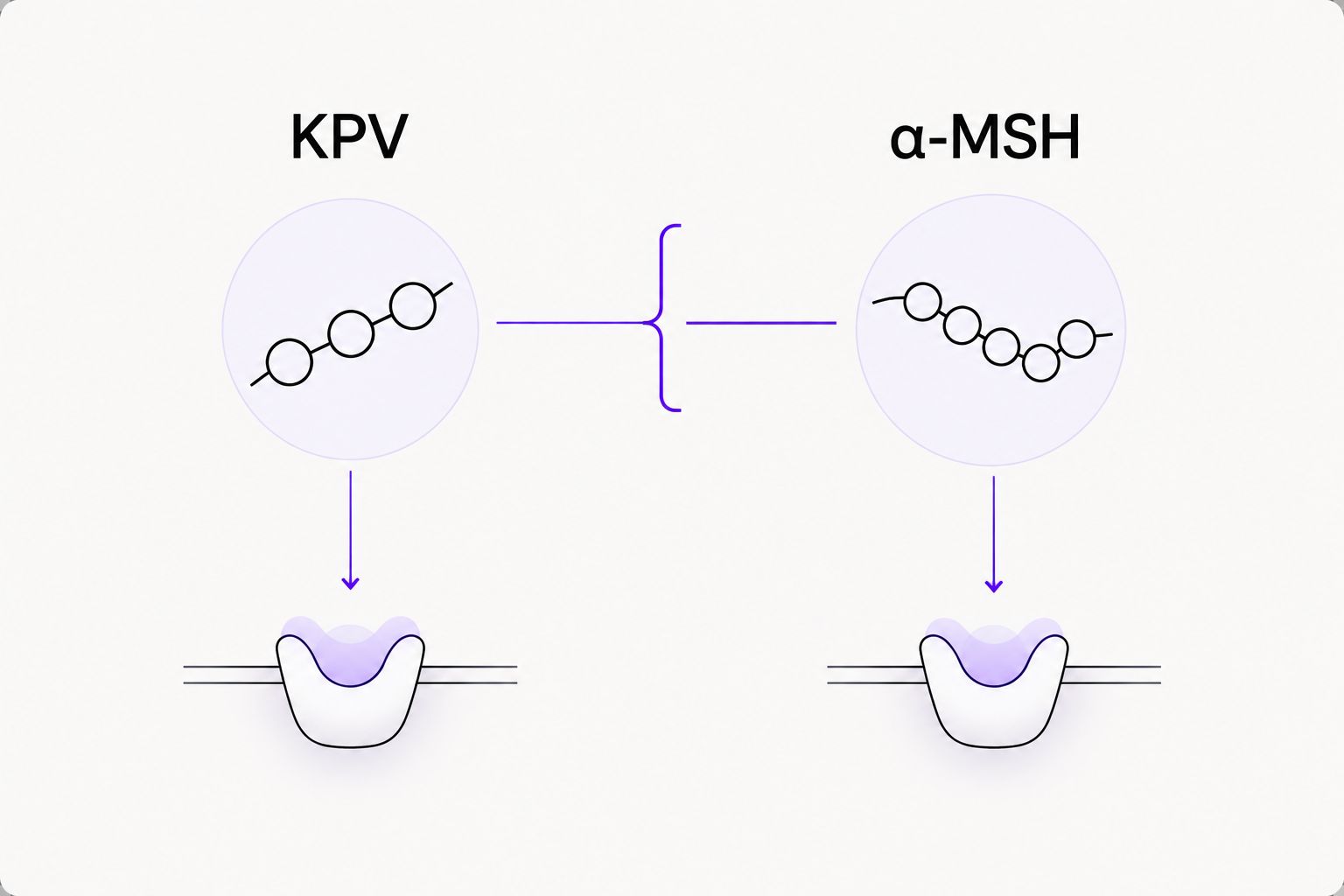
KPV contains 3 amino acid residues: lysine, proline, and valine. Its sequence corresponds to positions 11–13 of α-melanocyte-stimulating hormone, commonly abbreviated α-MSH, which contains 13 residues. The sequence relationship explains why the compounds appear together in the literature; it does not make their pharmacology interchangeable. 1
α-MSH belongs to the melanocortin signaling system. KPV retains only the C-terminal tripeptide sequence, not the complete molecular structure of α-MSH. Assigning the parent's receptor behavior to the fragment would therefore substitute a structural association for experimental evidence.
| Property | KPV | α-MSH |
|---|---|---|
| Sequence length | 3 residues | 13 residues |
| Structural relationship | C-terminal tripeptide sequence | Parent peptide containing that sequence |
| Mechanistic emphasis here | PepT1 uptake and intracellular inflammatory signaling | Melanocortin receptor pharmacology |
| Research advantage | A defined fragment for examining sequence-specific activity | A reference for parent-peptide signaling questions |
| Interpretive limitation | Fragment activity does not establish parent-like receptor engagement | Parent-peptide evidence cannot establish fragment behavior |
Non-interchangeability rule: KPV results belong to KPV; α-MSH results belong to α-MSH. Apply the same rule to terminal modifications and sequence variants. A related name is not sufficient evidence of chemical equivalence.
What PepT1 uptake establishes
Transport provides access, not a complete mechanism
PepT1, encoded by SLC15A1, is a proton-coupled transporter associated with dipeptide and tripeptide uptake. In the KPV study, transporter-mediated entry provided a mechanistic connection between extracellular peptide and intracellular signaling changes in the examined epithelial systems. 1
That distinction changes how you read an experiment. Extracellular exposure alone does not establish intracellular access, and transporter expression alone does not establish that transport explains the entire response. Evidence linking uptake to activity is stronger than either observation in isolation.
Keep the causal chain separated
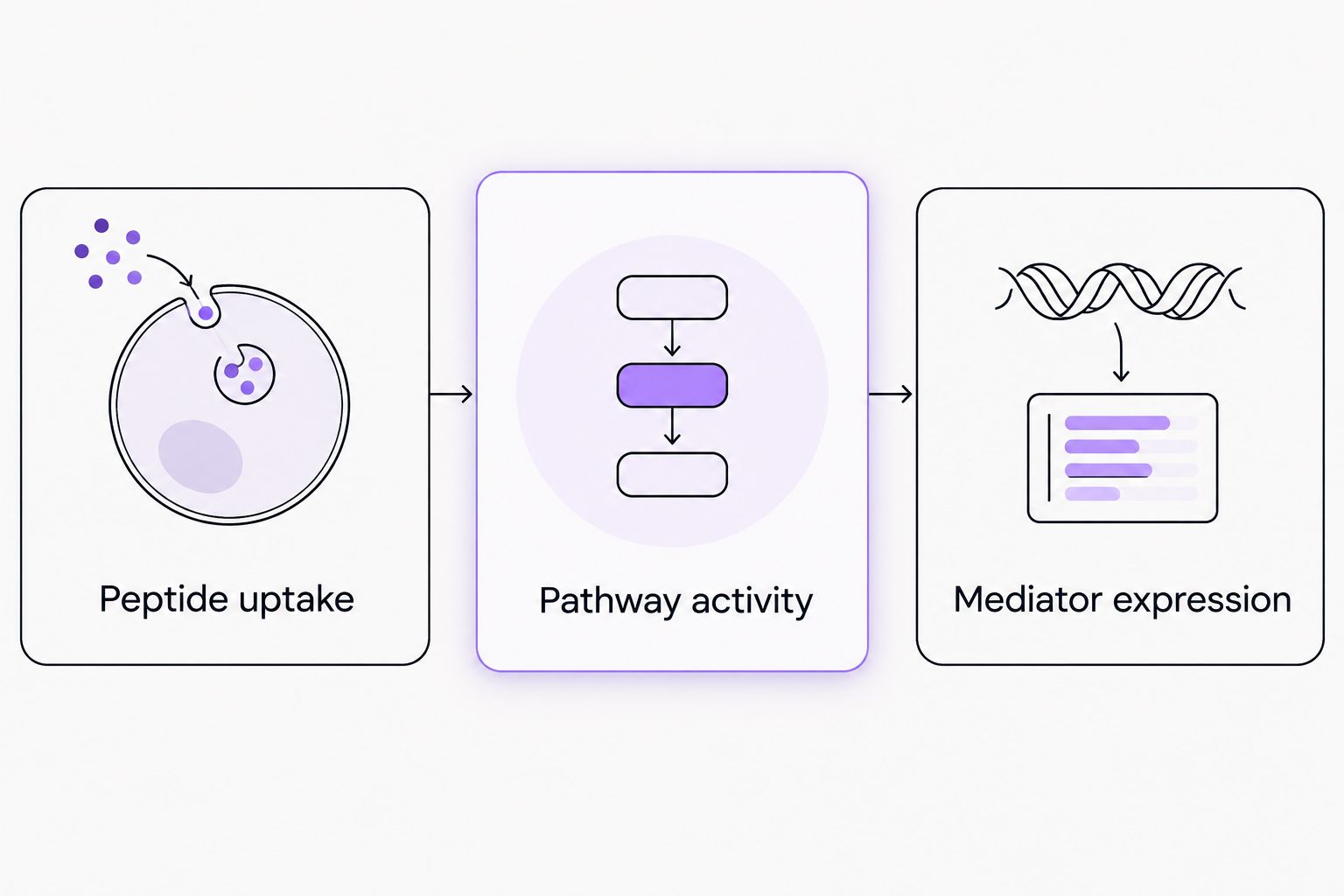
The published interpretation can be organized into three connected questions:
- Peptide uptake: Does the research system support entry of the identified peptide?
- Pathway activity: Does an intracellular signaling readout change?
- Mediator expression: Do downstream inflammatory measurements change consistently?
These are evidence categories, not a laboratory protocol. Their value is interpretive: they prevent a downstream measurement from being presented as proof of a specific initiating interaction.
The 2026 interpretation remains specific: PepT1-dependent uptake supports a transport-linked explanation in the reported system, not a universal explanation for every cell type. Establishing relevance elsewhere requires evidence from that research system rather than extrapolation from the intestinal-cell findings.
What NF-κB measurements establish
A pathway readout is not a binding assay
NF-κB is a family of transcription factors involved in inflammatory gene regulation. Its activity is commonly evaluated through measurements such as nuclear localization, DNA-binding activity, or transcriptional reporters. These measurements describe different aspects of pathway behavior; they are not interchangeable measurements of peptide binding.
The KPV literature reports inhibition of NF-κB-associated inflammatory signaling in the cellular systems examined. That supports a pathway-level statement. It does not, by itself, identify an enzyme, receptor, or transcription-factor subunit as a direct KPV binding partner. 1
Downstream markers need their own interpretation
A change in inflammatory mediator expression strengthens a signaling interpretation when it agrees with other measurements. Alone, however, lower expression does not locate the molecular interaction responsible for the change. General loss of cellular function can also reduce a biological readout, which is why viability and assay-performance controls matter.
Use precise wording in your research summary:
- Supported: KPV reduced the measured inflammatory signaling response in the specified cellular system.
- Not established by that result: KPV directly binds NF-κB.
- Not established by that result: KPV blocks every inflammatory pathway.
For a 2026 evidence table, record the actual endpoint beside each claim. “NF-κB inhibition” is too broad when the underlying result measures only one aspect of pathway activation.
Why KPV findings vary
Differences between research systems affect the interpretation of a KPV result. These factors identify where comparisons need qualification; they do not provide a basis for assigning an effect size.
- Transporter context: PepT1-mediated uptake is central to the cited epithelial-cell evidence. A different transport context requires separate support.
- Cellular background: Receptor expression, transcriptional state, and baseline inflammatory signaling differ across cellular systems.
- Inflammatory stimulus: A result obtained under one signaling challenge does not establish an identical response under another.
- Endpoint selection: Transport measurements, transcriptional reporters, and mediator expression capture different parts of the causal chain.
- Material identity: Sequence, terminal chemistry, and degradation products determine what material the experiment actually evaluates.
- Assay interference: A reduced signal needs interpretation alongside controls that distinguish biological activity from measurement artifacts.
Compare like endpoints before comparing conclusions. Two papers can use the same pathway name while measuring different biological events. Your summary should preserve that distinction rather than combine unlike results into a single mechanism claim.
How analytical documentation supports interpretation
Identity and purity answer different questions
Chromatographic purity describes the distribution of detected components under a specified analytical method. Mass spectrometry supports molecular identity through measured mass information. Neither measurement alone establishes receptor selectivity, intracellular transport, or biological activity.
A high chromatographic area percentage is therefore not a mechanism result. It also does not automatically establish peptide content by mass, because water, counterions, and other components can affect the relationship between weighed material and peptide quantity.
| Documentation element | What it supports | What it does not establish |
|---|---|---|
| Sequence and terminal chemistry | Definition of the intended molecular entity | Biological equivalence to related peptides |
| Chromatogram and method | Separation and detected-component profile | Complete impurity identification |
| Mass spectrum | Mass-based identity assessment | Full structural confirmation in isolation |
| Batch identifier | Traceability between material and records | Independent analytical verification |
| Stability information | Interpretation of material integrity under documented conditions | Stability under every laboratory condition |
Connect the result to the actual batch
For research-grade chemicals, 4-Amino-Labs serves laboratory buyers rather than consumer-use applications. When evaluating KPV sourcing, use the KPV research supplier guide as a documentation-oriented starting point, not as evidence that any supplied batch reproduces a published biological result.
4-Amino-Labs research materials require the same distinction between supplier information and experiment-specific evidence as materials from any source. Supplier identity cannot replace analytical review.
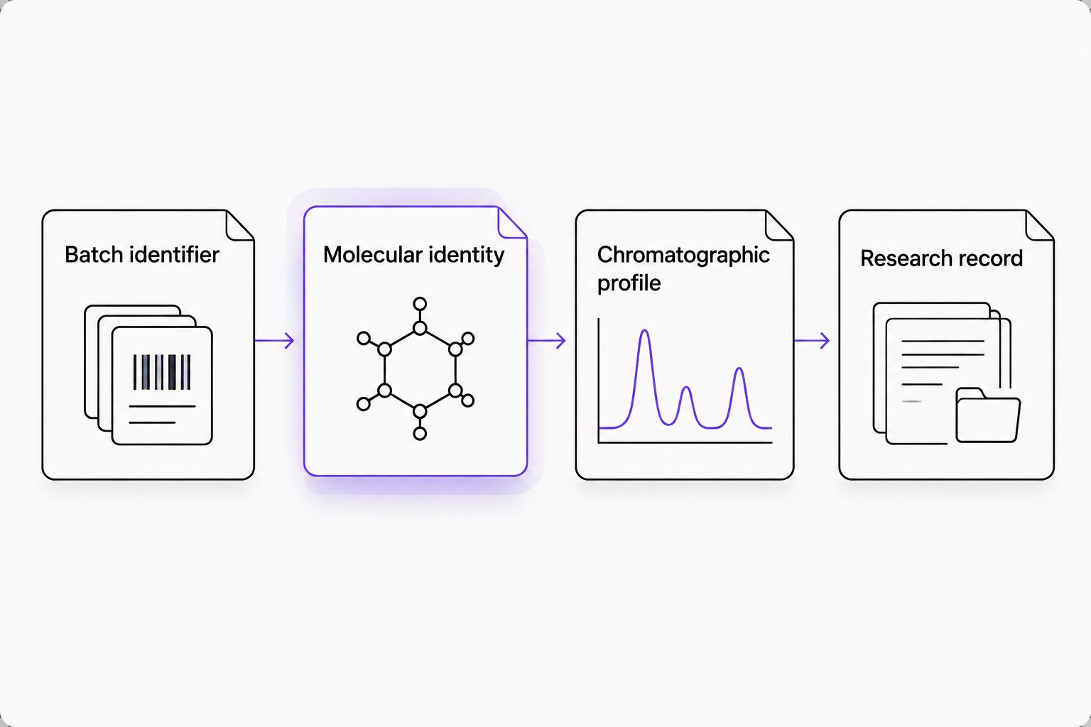
In a 2026 research record, preserve the relationship between the batch identifier, analytical files, and experimental observations. A generic certificate detached from the material cannot establish that relationship.
Related mechanism questions
Is KPV a melanocortin receptor agonist like α-MSH?
KPV's 3-residue sequence does not establish α-MSH-like melanocortin receptor agonism. The transport-linked inflammatory signaling evidence should be described on its own terms rather than assigned the parent's receptor mechanism. 1
Does PepT1 expression predict a KPV response?
PepT1 expression alone does not predict a KPV signaling response. Transporter abundance, functional uptake, and downstream signaling measurements are separate evidence categories; expression data cannot substitute for all three.
Does reduced NF-κB activity establish direct inhibition?
Reduced NF-κB activity establishes a pathway-associated response, not direct molecular binding. A direct-target claim requires evidence capable of distinguishing physical interaction from an upstream or indirect effect.
Limitations of the evidence
The principal mechanistic source discussed here is a published study containing intestinal epithelial cell experiments. This guide addresses that cellular evidence without extending findings across unrelated systems or describing administration, dosing, or organism-level applications. 1
The strongest feature of the evidence is its connection between peptide transport and inflammatory signaling. Its boundary is equally important: a transport-linked cellular response is not a complete molecular target map.
For your 2026 interpretation, keep these limits explicit:
- A defined experimental system establishes context, not universal activity.
- Shared sequence does not establish shared receptor occupancy or affinity.
- Reduced inflammatory markers do not establish selectivity across unrelated pathways.
- Analytical identity does not establish biological potency.
Describe what was measured before naming what you think caused it. This keeps the conclusion proportional to the evidence and makes later comparisons more reliable.
FAQ
What is the KPV peptide mechanism in inflammatory signaling research?
The KPV peptide mechanism links PepT1-mediated uptake with reduced NF-κB-associated inflammatory signaling in published intestinal epithelial cell experiments. That finding supports a transport-linked cellular mechanism, not a universal receptor assignment.
What amino acids make up KPV?
KPV consists of lysine, proline, and valine, a sequence of 3 amino acid residues. It corresponds to the C-terminal sequence at positions 11–13 of α-MSH.
Is KPV the same research compound as α-MSH?
KPV and α-MSH are different molecular entities. KPV contains 3 residues, whereas α-MSH contains 13 residues; their shared sequence does not establish equivalent receptor pharmacology.
Is PepT1 a receptor for inflammatory signaling?
PepT1 is a peptide transporter, not an inflammatory signaling receptor. Its relevance to the cited KPV research concerns cellular uptake and the connection between uptake and downstream activity.
Does KPV directly bind NF-κB?
A reduction in NF-κB-associated activity does not establish direct KPV binding to NF-κB. Direct-target claims require evidence beyond pathway or inflammatory-marker measurements.
Does a peptide purity result establish KPV biological activity?
A peptide purity result does not establish KPV biological activity. Chromatographic characterization, mass-based identity assessment, and biological measurements answer different questions.
Does this guide provide instructions for using KPV?
This guide provides no administration, dosing, or product preparation instructions. Its scope is molecular identity, cellular signaling evidence, analytical interpretation, and research documentation.
One last thing
The most useful correction is terminological: a transporter is not a receptor, and a pathway response is not a binding result. Apply those distinctions to your evidence table before adding more mechanistic detail. They prevent the central KPV finding from becoming a broader claim than the experiment supports.
References
- Dalmasso G, Charrier-Hisamuddin L, Nguyen HTT, Yan Y, Sitaraman S, Merlin D. PepT1-mediated tripeptide KPV uptake reduces intestinal inflammation. Gastroenterology. 2008;134(1):166–178. Scope cited here: peptide uptake and inflammatory signaling in the paper's cellular experiments.
Related guides
- Connecting batch numbers to laboratory inventory tracking
- Connecting research chemical purchases to a laboratory notebook
- Peptide research for biotech startups
Research use only. This content concerns laboratory research materials and scientific interpretation only. It is not medical advice and is not intended to diagnose, treat, cure, or prevent disease. No human or animal use is described or recommended.